Salmonella Test Results Hold Promise For Non-Invasive Cancer Treatment
An infamous food-borne bacteria's ability to penetrate cells has led to treatments that actively target and treat prostate cancer in new studies.
An international team comprising engineers, mathematicians and doctors has applied a technique used for detecting damage in underwater marine structures to identify cancerous cells in breast cancer histopathology images.
Their multidisciplinary breakthrough, which has the potential to automate the screening of images and improve the detection rate, has been published in leading journal, PLOS ONE.
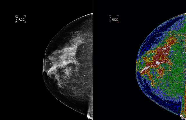
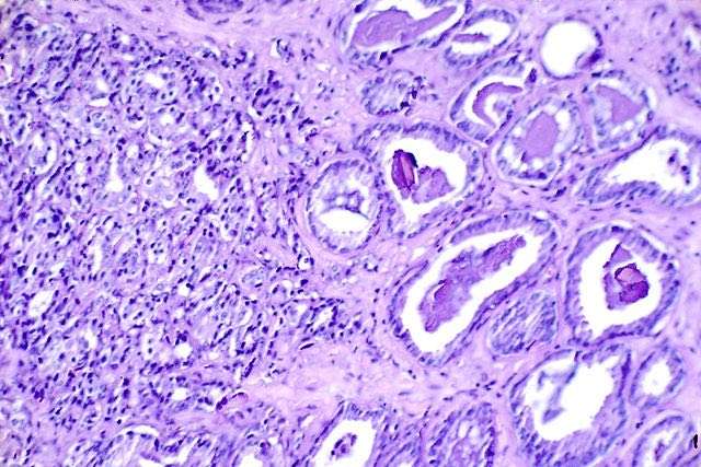
Breast cancer is the most prevalent form of cancer for women worldwide. Current breast cancer clinical practice and treatment mainly relies on the evaluation of the disease's prognosis using the Bloom-Richardson grading system. The necessary scoring is based on a pathologist's visual examination of a tissue biopsy specimen under microscope, but different pathologists may assign different grades to the same specimens.
However, the advent of digital pathology and fast digital slide scanners has opened the possibility of automating the prognosis by applying image-processing methods. While this undoubtedly represents progress, image-processing methods have struggled to analyse high-grade breast cancer cells as these cells are often clustered together and have vague boundaries, which makes successful detection extremely challenging.
But the new method has seemingly overcome that task, according to Assistant Professor in Civil Engineering at Trinity College Dublin, Bidisha Ghosh. She said: "This unique research group could draw on a broad and deep knowledge base. Experts in numerical methods and image-processing liaised with medical pathologists, who were able to offer expert insight and could tell us precisely what information was of value to them. It is an excellent example of how multidisciplinary research collaborations can address important societal issues."
Professor Joy John Mammen, Head of Department of Transfusion Medicine & Immunohaematology from the Christian Medical College, Vellore, India, said: "Detection of cancerous nuclei in high-grade breast cancer images is quite challenging and this work may be considered as a first step towards automating the prognosis."
This technique was developed in conjunction with mathematicians in Madras Christian College, India. Lead author, Dr Maqlin Paramanandam, said: "The potential for this technology is very exciting and we are delighted that this international and inter-disciplinary team has worked so well at tackling a real bottle-neck in automating the diagnosis of breast cancer using histopathology images."
Dr Michael O'Byrne, who also worked in University College Cork during this project, added: "Coming from a civil engineering background where most of our image-processing tools were designed to assess structural damage, it was nice to discover some cross-over applications and find areas where we could lend our expertise. We all found it particularly rewarding to contribute towards breast cancer research."
(Source: Trinity College Dublin)
Multiply The Good: Click To Share – Photo by NASA Goddard Photo and Video, CC license


Be the first to comment