Plant Toxins Fatal to Sugarcane Hailed as the 'New Weapon' Antibiotic in Fight Against Bacteria
The threat of antibiotic-resistant bacterial infections has been growing across human civilization for years.

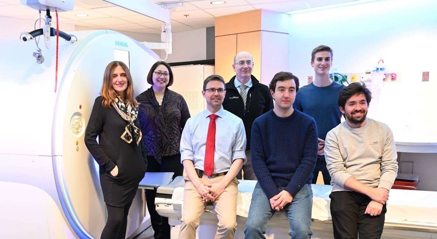
Researchers at MIT (Massachusetts Institute of Technology) have developed a new AI deep learning model that can predict lung cancer risk up to six years in advance through a single low-dose CT scan.
Lung cancer is the deadliest cancer in the world-resulting in more deaths than the next three cancers combined. It is also extremely difficult for humans to find the disease early by looking at scans.
Current lung cancer prediction models require a combination of demographic information, clinical risk factors, and radiologic annotations, whereas the model called 'Sybil' is designed to use a single low dose chest scan to predict the risk of lung cancers occurring 1-6 years after a screening.
Peter Mikhael, co-first-author and PhD student at MIT likened the overall process of lung cancer screening to "trying to find a needle in a haystack."
However, working with a diverse set of scans from two hospitals and the National Lung Cancer Screening Trial, the study showed Sybil was able to forecast both short-term and long-term lung cancer risk, earning C-indices scores ranging from 0.75 to 0.80. Values over 0.8 indicate a strong model.
When predicting cancer risk one year in advance, the model was even more successful: they obtained between 0.86 to 0.94 on a ROC-AUC probability curve (considered excellent for AUC values with 1.00 being the highest possible score).
The imaging data used to train Sybil was largely absent of any signs of cancer because early-stage lung cancer occupies small portions of the lung-just a fraction of the hundreds of thousands of pixels making up each CT scan.
Denser portions of lung tissue known as lung nodules have the potential to be cancerous, but most are not and are, instead, healed infections or airborne irritants.
Co-author Jeremy Wohlwend was surprised by how highly Sybil scored, despite the lack of any visible cancer.
"We found that while we as humans couldn't quite see where the cancer was, the model could still have some predictive power as to which lung would eventually develop cancer."
Professor Regina Barzilay led the research team at the Jameel Clinic at MIT, in partnership with Mass General Cancer Center and Chang Gung Memorial Hospital in Taiwan, which published the study in the Journal of Clinical Oncology.
This model aims to bring the research community one step closer to outgrowing legacy systems in the healthcare industry and help better treat current and future patients.
SHARE This Medical Breakthrough With Friends on Social Media…
Be the first to comment